|
|
Imaging
buy prednisolone 5mg tablets uk cheap prednisolone click buy prednisolone How to Take Amoxicillin buy antibiotics online link amoxicillin online amitriptyline 25mg amitriptyline online abortion in philippines abortion pill ph open abortion pill philippines usa buy abortion pill purchase abortion pill online where can i buy duloxetine duloxetine buy online uk tadalafil generico sandoz cialis generico 10 mg prezzo link
Team Members:
- Mr. Javed Iqbal (Electron Microscopist)
|
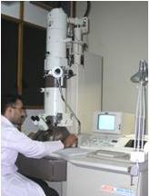
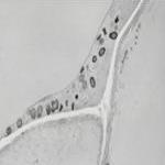
Transmission electron microscopy:
The transmission electron microscope (TEM) works on the same basic principles as the light microscope but uses electron beam instead of light. Light microscope is limited by the wavelength of light but a TEM uses electron as “light source” and their much lower wavelength makes it possible to get a resolution of a few angstrom (10-10 m). At the bottom of the microscope the un-scattered electrons hit the fluorescent screen, which gives rise to a "shadow image" of the specimen with its different parts displayed in varied darkness according to their electron density. The images are photographically recorded by exposing a photographic film. The possibility for high magnifications has made the TEM a valuable tool in both medical, biological and materials research.
|
 |
|
At NIBGE, this state of the art technique is not only been handled with great care but has been utilized regularly in numerous studies related to different topics. Currently this is the only instrument providing services, not only to the scientists / scholars of NIBGE but also to the other scientific Institutes, organizations and Universities of the Pakistan in this regards the Higher education commission of Pakistan (HEC) has made a great contribution through its “Access to Scientific Instrumentation Project” extending this facility to all over the country.
|
 |
|
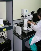
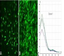
Confocal Laser Scanning Microscopy
Laser Scanning Confocal Microscopy has become an invaluable tool for a wide range of investigations in the biological and medical sciences as well as in materials science for imaging thin optical sections in living and fixed specimens ranging in thickness up to 100 micrometers. Confocal microscopy permits non-destructive imaging of translucent specimens or opaque surfaces, elimination of background information away from focal plane, collection of serial optical sections from thick specimens and depth of field control. Modern instrument is equipped with laser systems controlled by high-speed acousto-optic tunable filters (AOTFs), coupled with photomultipliers, beam scanning assembly, electronic detectors and computers for image display, processing, output and storage. The application of a wide array of new synthetic and naturally occurring fluorochromes has made it possible to identify cells and sub-microscopic cellular components with a high degree of specificity under Confocal Laser Scanning Microscope. Through the use of multiply-labeled specimens, different probes can simultaneously identify several target molecules, both in fixed specimens and living cells as well as tissues. Many fluorescent probes are constructed around synthetic aromatic organic chemicals, designed to bind with a biological macromolecule (for example, a protein or nucleic acid) or to localize within a specific structural region (such as the cytoskeleton, mitochondria, Golgi apparatus, endoplasmic reticulum, and nucleus). Other probes are employed to monitor dynamic processes and localized environmental variables (including concentrations of inorganic metallic ions, pH, reactive oxygen species, and membrane potential). Fluorescent dyes are also useful in monitoring cellular integrity (live versus dead and apoptosis), endocytosis, exocytosis, membrane fluidity, protein trafficking, signal transduction, and enzymatic activity. In addition, fluorescent probes have been widely applied to genetic mapping and chromosome analysis in the field of molecular genetics. |
|
?
|